A brain aneurysm diagnosis can be terrifying. Even though a ruptured aneurysm is somewhat infrequent, it can still signify a very serious illness that can lead to disability and even death.
Here is an overview of what a brain aneurysm is and what the surgical treatment entails. Information about this condition can help ease fears and bring a sense of support during a challenging time.
What is a brain aneurysm?
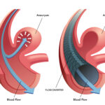
Otherwise known as a cerebral aneurysm, this condition occurs when there is a bulging spot on the wall of a brain artery, which can be likened to a thin balloon.
As time goes on, the blood flow within the artery will hit against the thin portion of the wall and aneurysms can form, causing significant wear on the arteries.
Once the artery wall becomes thinner, the blood flow causes the weakened wall to swell.
The pressure can cause the aneurysm to burst and blood will flow around the brain. If this happens, surgery is often needed to fix it.
What are the symptoms?
Brain aneurysms that are unruptured often will not cause any symptoms.
But, if the aneurysm is large enough, it can sometimes press on the brain or nerves which can cause neurological symptoms, including dilated pupils, blurred or double vision, pain above and behind the eye, nausea/vomiting, stiff neck, loss of consciousness, difficulty speaking, localized headache, weakness, or numbness.
What does the brain aneurysm surgery involve?
There are two common surgical approaches to repairing an aneurysm. The first method is when the doctor clips the aneurysm, which is completed through an open craniotomy.
Here, the physician creates a hole in the skull to attend to the area where the aneurysm is located.
The other way it can be treated is through endovascular repair, which is a less invasive method that involves inserting a coil or stent.
Patients who undergo the aneurysm clipping will be put under general anesthesia with a breathing tube.
Then the scalp, skull, and coverings of the brain are opened. A metal clip is inserted at the neck of the aneurysm to prevent it from bursting.
Patients who have the endovascular repair will either have general anesthesia or a medicine to sedate them without putting them to sleep.
A catheter is guided through the groin to an artery, then the blood vessel where the aneurysm is located.
A contrast dye is injected through the catheter which allows the physician to look at the arteries and the aneurysm on a monitor.
Once it is located, thin metal wires are put into the aneurysm, which then coil into a mesh ball.
Sometimes, mesh tubes are also put in to keep the coils secure. If blood clots form around the coil, it will prevent the aneurysm from bursting.
After the surgery is completed and deemed successful, it isn’t common to have a bleeding aneurysm again in the same area.
Patients will likely need to be seen by a doctor every year to discuss progress and prognosis, but most of the time these surgical repairs can prevent further brain aneurysms.


